
Aggressive T10 posterior-element lesion — recognising cord compression as an urgent MRI finding
Key images
Clinical context
An adult presented with severe lower back pain radiating to both lower limbs. Multiplanar, multisequence MRI of the thoracic spine was performed without intravenous contrast.
Key finding
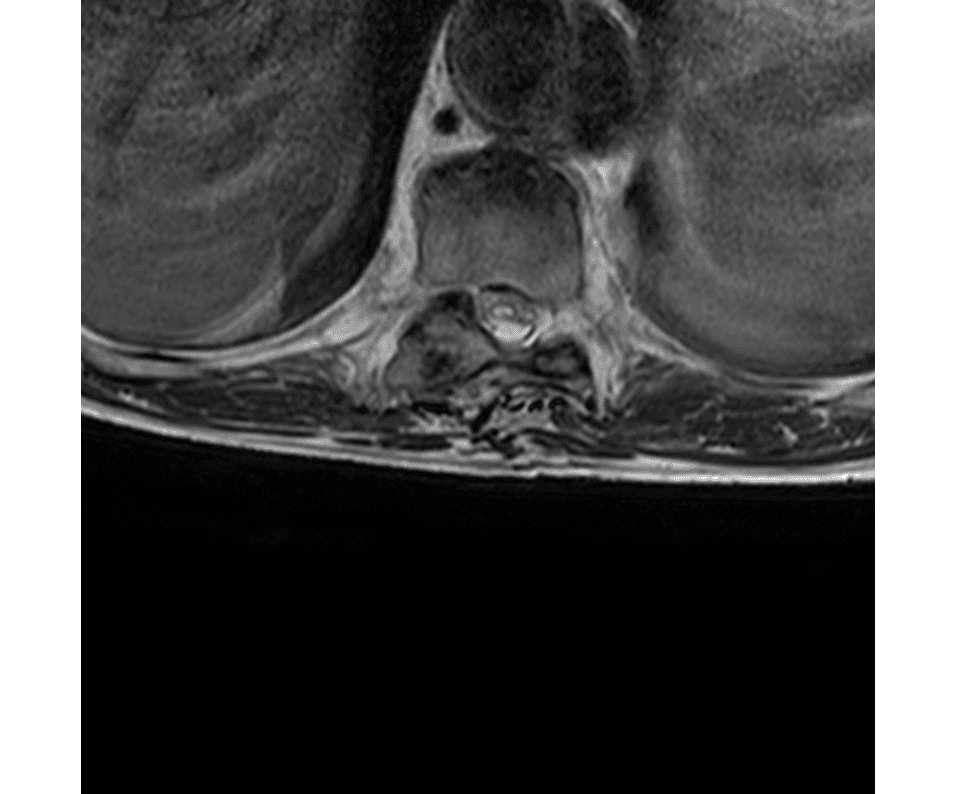
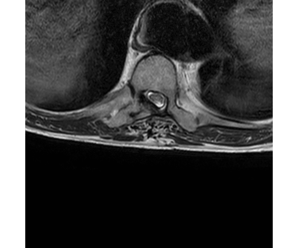
An expansile lesion involves the right posterior elements of T10, extending into the right costovertebral joint and the expanded proximal right 10th rib, with an extraosseous soft-tissue component and right paraspinal extension. Epidural extension produces mild-to-moderate central canal stenosis with anterior displacement of the thecal sac and cord compression. A focal T2/STIR-hyperintense intramedullary signal abnormality is centred at T10–T11, measuring approximately 2.5 cm craniocaudally, consistent with compressive myelomalacic change/cord oedema. No additional focal thoracic osseous lesion or significant disc disease was identified.
Teaching point
An expansile lesion of the posterior elements with costovertebral, rib and paraspinal extension plus an extraosseous soft-tissue mass has an aggressive appearance and should prompt consideration of primary or metastatic osseous neoplasm. The critical, time-sensitive observation is epidural extension with canal stenosis and intramedullary T2 hyperintensity — cord compression with cord signal change is an urgent finding. It mandates contrast-enhanced MRI, CT for osseous characterisation, tissue diagnosis, and immediate neurosurgical/orthopaedic-oncology involvement.
Teaching case shown for educational purposes. Anonymized; no identifiable patient data. A radiology report is a specialist opinion and must be interpreted with the full clinical context by the treating physician.